
Kidney Cysts: Simple vs. Complex Renal Cyst Evaluation — Reassurance, Precision Diagnostics & Definitive Action
Discovering a renal cyst on an incidental ultrasound or CT scan is extremely common — in fact, over 50% of adults over the age of 50 develop simple cysts, and the vast majority are 100% benign. However, precision imaging and expert Bosniak stratification are essential to differentiate harmless fluid sacs from complex, septated, or suspicious cystic lesions that demand active oncologic management.
18+ Yrs
Surgical Leadership
12,000+
Urology Surgeries
Daycare MIST
Rezūm, UroLift, iTind
Clinical Pathological Comparison
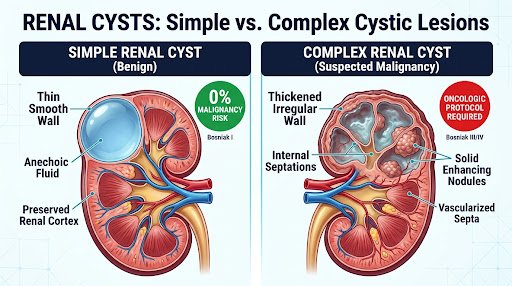
Simple Cyst (Benign)
Thin, smooth wall, purely liquid, no vascular uptake. 0% cancer risk.
Complex Cyst (Suspicious)
Thick irregular walls, septa, or solid nodules requiring urgent scan triage.
Simple vs. Complex Renal Cysts: What Does Your Scan Report Actually Mean?
When an ultrasound report notes a "Cystic lesion in the kidney", understanding radiological descriptors empowers you to know whether observation or surgical consult is appropriate.
0% Risk
Simple Renal Cyst (Harmless & Benign)
A thin-walled sac filled with pure serous fluid. Represents normal involutional aging of the kidney tubules; never converts into cancer.
Anechoic Fluid Matrix:
Pure, clear serous fluid with no internal echoes or debris on ultrasound.
Hair-thin Capsule & Posterior Enhancement:
Smooth, razor-thin wall boundary without calcified plaques or irregular humps.
Zero Contrast Enhancement:
No vascular blood flow pickup on contrast-enhanced CT or MRI scans.
Clinical Symptom Profile:
98% asymptomatic; incidental finding during routine full-body checkup.
5% to >90% Risk
Complex Cystic Lesion (Oncologic Evaluation)
A fluid-containing cavity featuring internal structural irregularities, partitioned chambers (septations), or tissue nodules that require dedicated oncologic review.
Thickened or Nodular Walls:
Coarse, irregular perimeter contours that deviate from a smooth fluid sphere.
Internal Septations (Chambers):
Multiple fibrous dividing walls within the cyst, especially if >1mm or enhancing.
True Contrast Enhancement (CT/MRI):
Hounsfield unit increase (>15-20 HU) post-contrast, indicating neo-vascularization.
Potential Red Flag Signs:
Microscopic or visible blood in urine (hematuria), persistent flank fullness.
The Bosniak Classification System Demystified
The Bosniak Classification System Demystified
Urologists and uro-radiologists utilize the internationally validated Bosniak Classification (updated to v2019) on contrast-enhanced CT or MRI to determine whether a cyst is safe to ignore, requires surveillance, or necessitates nephron-sparing excision.
Category I
Simple Benign Renal Cyst
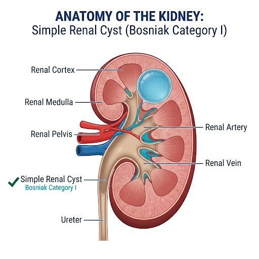
Key Diagnostic Features
- Hairline paper-thin smooth wall, hair-like outer capsule
- Pure anechoic clear fluid matrix (water attenuation)
- No internal septa, calcification, or vascular enhancement
Category II
Minimally Complex Benign
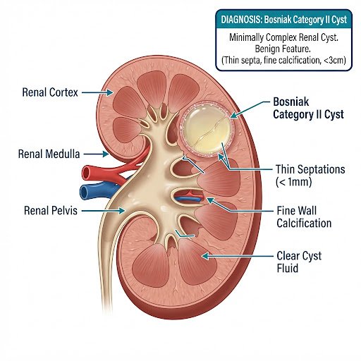
Key Diagnostic Features
- Few hairline thin internal septa (<1 mm) without nodularity
- Fine wall or septal calcification specks
- High-attenuation non-enhancing fluid sac (<3 cm)
Category IIF
Follow-up / Surveillance
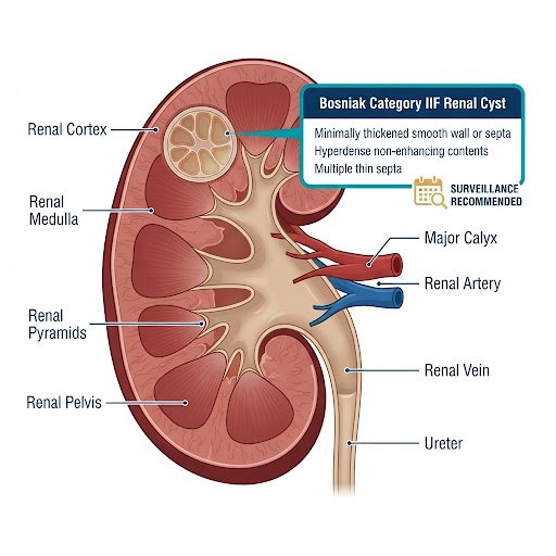
Key Diagnostic Features
- Multiple hairline thin septa or minimal smooth wall thickening
- Nodular or thick calcification without measurable uptake
- Totally intrarenal hyperdense non-enhancing cyst (>3 cm)
Category III
Indeterminate Complex Cyst
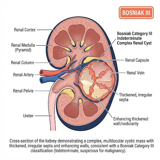
Key Diagnostic Features
- Thick, irregular or multi-layered septa (>2 mm)
- Measurable contrast enhancement of wall or internal septa
- Multilocular complex compartments requiring surgical review
Category IV
Malignant Cystic Carcinoma
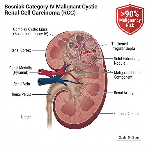
Key Diagnostic Features
- Solid enhancing nodular components adjacent to wall/septa
- Thickened, vascularized, irregular invasive septa
- Unequivocal cystic renal cell carcinoma (RCC) hallmarks
Diagnostic Algorithm
Rajshree Apollo Uro-Oncology Protocol
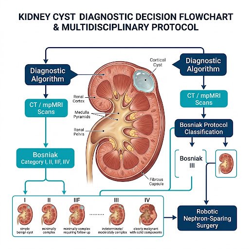
Multi-Disciplinary Decision Protocol
- Tri-phase Dual-Energy CT & High-Tesla MRI confirmation
- Differentiates surgical malignancy from benign cyst mimics
- Da Vinci Xi robotic console preserves >90% functioning parenchyma
"Over 95% of simple cysts never need surgery."
Patients often arrive distressed after reading "Cyst discovered on kidney" on a sonography sheet. Dr. Chipde emphasizes non-invasive observation for uncomplicated simple cysts. Intervention is strictly reserved for symptomatic or mechanically obstructive cysts.
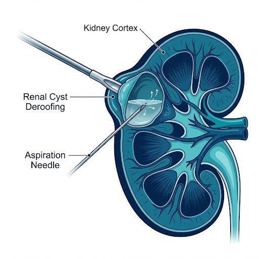
5 Specific Clinical Scenarios When a Benign Cyst Requires Treatment
If a simple cyst causes structural compression or clinical distress, Dr. Chipde provides minimally invasive, scar-less interventions.
01
Giant Cortical Cysts (>7 to 10 cm) Causing Mass Effect
Massive cysts that stretch the renal capsule or compress the digestive viscera, leading to early satiety or abdominal fullness.
02
Pelviureteric Junction (PUJ) Compression & Hydronephrosis
Cysts situated near the renal hilum that pinch the ureter or renal pelvis, impeding urinary outflow and causing kidney swelling.
03
Chronic Refractory Flank or Lower Back Pain
Persistent dull aching in the costovertebral angle that does not respond to conservative therapy and correlates directly with cyst location.
04
Secondary Cyst Infection, Hemorrhage, or Abscess
Spontaneous bleeding into the cyst cavity causing acute stabbing pain, or bacterial colonization producing fever and infected pyocells.
05
Renovascular Hypertension (Page Kidney Phenomenon)
Subcapsular cysts exerting localized pressure on the main renal artery branches, triggering severe secondary high blood pressure via renin secretion.
The 5-Step Diagnostic & Management Pathway at Rajshree Apollo Hospitals
From incidental sonography doubt to decisive, organ-preserving resolution under Dr. Saurabh Sudhir Chipde.
01
Multiphase Tri-Phase CT / High-Tesla MRI
High-resolution non-contrast, corticomedullary, and nephrographic contrast phases to accurately quantify Hounsfield Units (20 HU enhancement).
02
Formal Bosniak Risk Stratification
Senior uro-oncologist reading to assign categorical score (I, II, IIF, III, or IV) eliminating false alarms and unnecessary invasive procedures.
03
Renal Functional & Nuclear Scan (DTPA/EC)
When a large or hilar cyst borders the renal pelvis, split renal function tests confirm if glomerular clearance or outflow is compromised.
04
Individualized Surveillance vs. Intervention
Patients with Category IIF join a low-radiation surveillance registry. Category I & II receive immediate clinical clearance.
05
Robotic Nephron-Sparing Console Precision
If Bosniak III/IV surgery is mandated, Da Vinci Xi robotic partial nephrectomy excises the cyst margins cleanly while saving 90%+ of normal kidney nephrons.
Frequently Asked Questions on Kidney Cysts & Bosniak Lesions
Evidence-based explanations to dispel misconceptions and assist in your healthcare decisions.
Do simple kidney cysts turn into cancer over time?
No. True simple cysts (Bosniak Category I) do not undergo malignant degeneration. They are benign fluid collections that arise from dilated renal tubules as a normal aging phenomenon. Once definitively proven to be a simple thin-walled cyst by ultrasound or contrast CT, it will not transform into kidney cancer. Only complex cysts with pre-existing suspicious features (thick septa, solid nodules) harbor cancer risk.
How often do I need follow-up scans for a Bosniak IIF cyst?
Bosniak IIF lesions ("F" stands for Follow-up) carry an estimated ~5% risk of harboring an indolent cystic neoplasm. European and American urological protocols mandate contrast-enhanced imaging (Dual-Energy CT or High-Tesla MRI to spare radiation) at 6 months, 12 months, and annually for up to 3 to 5 years. If the septations and walls remain strictly stable with zero progressive enhancement, the cyst is considered clinically benign.
Can a simple kidney cyst cause high blood pressure or back pain?
In rare cases, yes. While the majority are completely asymptomatic, a very large simple cyst (>7-10 cm) or a cyst located strategically at the kidney's vascular hilum can mechanically compress the main renal artery or stretch the capsule. This pressure can trigger renin release resulting in secondary renovascular hypertension (Page kidney phenomenon) or dull costovertebral flank discomfort. Decompressing the cyst through minimally invasive unroofing frequently normalizes blood pressure and resolves the ache.
What is the difference between a simple kidney cyst and Polycystic Kidney Disease (ADPKD)?
They are entirely distinct conditions. A simple cyst is an isolated, sporadic, localized non-genetic finding occurring in 1 or 2 areas of otherwise completely healthy kidneys. Autosomal Dominant Polycystic Kidney Disease (ADPKD) is an inherited genetic mutation (PKD1/PKD2 genes) causing bilateral kidneys to progressively fill with hundreds of enlarging cysts over decades, causing renal enlargement and progressive renal insufficiency. Having one or two simple cysts does not mean you have ADPKD.
How does robotic nephron-sparing surgery protect my overall kidney function?
For suspicious Bosniak Category III or IV lesions, traditional surgery often removed the entire kidney (radical nephrectomy). Dr. Saurabh Sudhir Chipde utilizes the Da Vinci Xi 4-arm robotic system to perform Robotic Partial Nephrectomy. The robotic 3D-HD optics and EndoWrist articulation allow exact millimeter precision: clamping the tumor blood vessel with minimal ischemia time, excising purely the complex cystic capsule with oncologically negative margins, and suturing the renal parenchymal defect. This preserves maximum working nephrons, protecting you against long-term cardiovascular disease and chronic kidney disease.
Get Expert Bosniak Stratification & Reassurance Today
Bring your ultrasound, CT scan, or MRI films to Rajshree Apollo Hospitals, Indore. Experience clear explanations, nephron-preserving philosophy, and world-class urologic care.
Visit Dr. Chipde at Rajshree Apollo Hospitals Indore
Conveniently accessible in Vijay Nagar, Indore. Book an express in-clinic consultation or connect on WhatsApp.
Rajshree Apollo Hospitals, Indore
- Practice Address:
Rajshree Apollo Hospitals, Sector-D, Scheme No. 74C, Vijay Nagar, Indore, Madhya Pradesh 452010
- OPD Clinic Hours:
Monday to Saturday: 10:00 AM – 06:00 PM (Sunday by emergency appointment)
- Direct Phone & WhatsApp: