
Kidney Cancer (RCC) & Partial Nephrectomy — Precision Excision, Kidney Preservation & Definitive Cure
Discovering a renal mass on an incidental ultrasound or CT scan is an anxious moment — but today, kidney cancer is one of the most curable urological malignancies when detected early. Over 70% of kidney tumors are discovered incidentally at T1 stage (≤7 cm). Dr. Saurabh Sudhir Chipde specializes in da Vinci® Robotic Partial Nephrectomy (Nephron-Sparing Surgery), meticulously excising the localized tumor with negative margins while saving over 90% of healthy functional kidney nephrons — preventing long-term chronic kidney disease and avoiding lifelong dialysis.
18+ Yrs
Surgical Leadership
12,000+
Urology Surgeries
Daycare MIST
Rezūm, UroLift, iTind
Evidence-Based Protocol
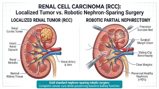
Zero Compromise on Oncologic Safety
Complete negative tumor margins with maximal nephron preservation, keeping baseline kidney function intact without sacrificing cancer cure rates.
What is Renal Cell Carcinoma (RCC)?
Renal Cell Carcinoma (RCC) originates primarily from the proximal tubular epithelial cells within the renal cortex. Accounting for over 90% of all primary kidney malignancies, modern urologic oncology categorizes RCC into well-defined histological variants that guide tailored robotic resection strategies.
Clear Cell RCC (ccRCC)
Characterized by clear cytoplasm rich in glycogen and lipids. While vascular, it is exceptionally responsive to localized robotic margin excision and systemic targeted therapies if advanced.
Prime Robotic Excision Target
Papillary RCC (pRCC)
Arranged in finger-like papillary projections. Divided into Type 1 (typically indolent, well-circumscribed) and Type 2 (higher nuclear grade). Often exhibits hypovascularity on contrast CT.
Prime Robotic Excision Target
Chromophobe RCC (chRCC)
Arises from the intercalated cells of the collecting ducts. Demonstrates prominent cell borders and halo appearance. Generally carries an outstanding long-term surgical prognosis.
Prime Robotic Excision Target
Oncocytoma & AML
Benign renal masses like Renal Oncocytoma and Angiomyolipoma (AML) frequently imitate RCC on standard ultrasonography, necessitating triple-phase CT assessment or robotic enucleation.
Prime Robotic Excision Target
The Modern "Incidentaloma" Era: Transforming Diagnosis into Cure
More than 70% of renal cell carcinomas are discovered incidentally during routine abdominal ultrasound or CT scans performed for unrelated concerns (such as fatty liver, dyspepsia, or kidney stones). Because these tumors are detected when asymptomatic and small (T1a/T1b), patients have the unique opportunity for a definitive cure through organ-sparing robotic surgery rather than radical removal.
Renal Cell Carcinoma TNM Staging & Surgical Pathway
Accurate anatomical and pathological staging governs the choice between Robotic Partial Nephrectomy, Radical Nephrectomy, and systemic precision immunotherapy.
T1a / T1b (≤ 7.0 cm)
Tumor is strictly confined within the renal capsule:
- T1a: Tumor ≤ 4.0 cm
- T1b: Tumor 4.1 cm to 7.0 cm
Gold Standard Treatment: Robotic Partial Nephrectomy (Nephron-Sparing). 5-Year Survival: 95%.
T2a / T2b (> 7.0 cm)
Tumor still strictly confined within kidney capsule but expanded:
- T2a: >7.0 cm but ≤ 10.0 cm
- T2b: >10.0 cm organ-confined
Surgical Selection: Extended Robotic Partial (if exophytic/favorable RENAL score) or Radical Nephrectomy. 5-Year: 80–85%.
T3 / N1 (Locally Advanced)
Extension into major veins or perinephric fat without breaching Gerota's fascia:
- T3a: Renal vein / perinephric fat
- T3b/c: Vena Cava extension
Procedure: Robotic Radical Nephrectomy with IVC Thrombectomy & Regional Lymphadenectomy.
T4 / M1 (Metastatic)
Tumor invades beyond Gerota’s fascia or has spread to distant organs (lungs, bone, liver):
- T4: Direct ipsilateral adrenal invasion
- M1: Distant organ metastasis
Multimodal Care: Cytoreductive Nephrectomy combined with modern dual-agent Immune Checkpoint Inhibitors.
Robotic Partial Nephrectomy vs. Traditional Radical Nephrectomy
International urological guidelines (EAU, AUA, USI) designate partial nephrectomy as the first-line treatment for localized renal masses because saving healthy parenchyma protects cardiovascular and overall longevity.
Clinical Parameter
da Vinci® Robotic Partial Nephrectomy
Traditional Radical Nephrectomy
Surgical Objective
Excision of localized tumor exclusively; preserves >90% healthy renal parenchymal filtration.
Complete removal of the entire kidney, surrounding fat, and often adjacent adrenal gland.
Renal Function (eGFR)
Preserves 85% to 95% of baseline filtration capacity; markedly minimizes risk of future dialysis.
Permanent 30% to 50% drop in renal clearance; elevated long-term cardiovascular mortality.
Tumor Size Eligibility
First-line Gold Standard for T1a (≤4 cm) and T1b (4–7 cm); selectively used in anatomically favorable T2.
Indicated for destructive tumors >7–10 cm or complete central invasion of renal hilum.
5-Year Oncologic Cure
>97% cancer-specific survival in localized lesions (identical to radical nephrectomy).
>97% cancer-specific survival (equal oncologic cure, but at expense of total organ loss).
Surgical Invasiveness
4 tiny keyhole ports (8 mm); EndoWrist 7 degrees of freedom dexterity; minimal blood loss (<100 ml).
Subcostal or flank incision (20–25 cm); cutting of abdominal wall muscles; higher post-op pain.
Hospital Inpatient Stay
48 hours (Accelerated ERAS recovery protocol).
5 to 7 days hospital stay due to open wound recovery.
Return to Daily Life
10 to 14 days for desk work and light routine activities.
6 to 8 weeks with substantial physical activity restrictions.
The 4-Step Robotic Partial Nephrectomy Surgical Technique
Under 10x high-definition 3D visualization, Dr. Saurabh Sudhir Chipde executes a four-phase operative sequence designed to guarantee complete tumor resection while strictly limiting parenchymal ischemic injury.
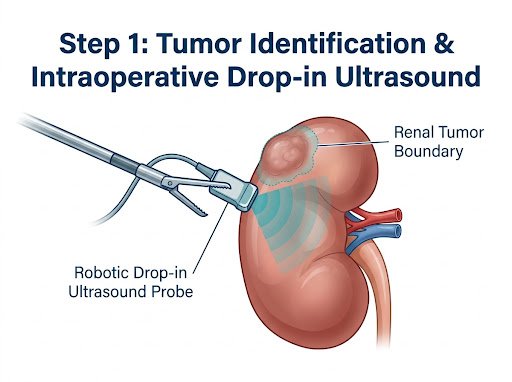
01
Tumor Identification & Drop-in US
Gerota’s fascia is incised and the renal mass exposed. A sterile robotic drop-in intraoperative ultrasound probe maps endophytic tumor depth and vascular proximity with millimetric accuracy.
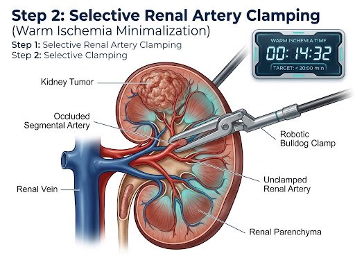
02
Selective Clamping (<20 min WIT)
Robotic bulldog clamps are selectively placed on branch renal arteries feeding the tumor. Warm Ischemia Time (WIT) is strictly controlled to <20 minutes, preventing reperfusion injury to remaining nephrons.
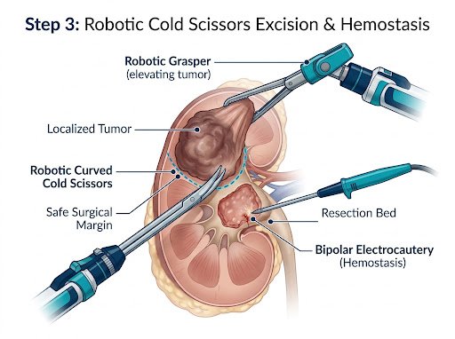
03
Cold Shears Precision Excision
The mass is excised using curved robotic cold scissors without excessive thermal spread. A uniform 1–2 mm normal parenchymal safety margin is carved out, ensuring oncologic negative margins.
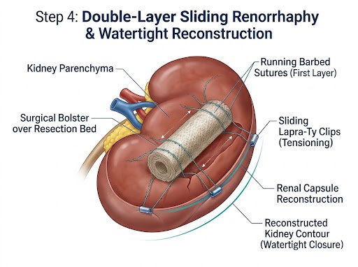
04
Double-Layer Sliding Renorrhaphy
Inner running barbed suture seals opened calyces and deep vessels. The outer parenchymal defect is secured with sliding Lapra-Ty clips over a surgical bolster, yielding complete, watertight hemostasis.
The Surgical Trifecta & Pentafecta Outcomes
In international uro-oncology, partial nephrectomy success is graded by strict composite benchmarks known as the Trifecta and Pentafecta. Dr. Saurabh Sudhir Chipde consistently exceeds these worldwide academic standards.
Criterion 01
Negative Margins
R0 complete oncologic clearance confirmed on frozen section & final pathology.
Criterion 02
WIT <20 Minutes
Zero thermal or ischemic necrosis of collateral functional glomeruli.
Criterion 03
No Complications
Zero major Clavien-Dindo Grade III-IV complications or readmissions.
Criterion 04
>90% GFR Saved
Long-term preservation of pre-operative baseline glomerular filtration.
Criterion 05
Recurrence-Free
Complete local disease control verified across multi-year CT follow-ups.
The 5-Step Multidisciplinary Care Pathway
From emergency report review to final histopathological surveillance, our integrated clinical pathway delivers world-class safety and zero delays.
01
Triple-Phase DECT / 3T mpMRI
Multi-phasic renal protocol imaging with 3D arterial reconstructions to evaluate precise tumor perfusion and vascular variants.
02
RENAL Nephrometry Scoring
Standardized anatomic grading of radius, exophytic properties, nearness to hilum, and polarity to optimize operative planning.
03
da Vinci Xi Robotic Excision
High-definition 3D surgery with selective arterial clamping and zero-tension sliding renorrhaphy under continuous hemodynamics.
04
Accelerated ERAS Recovery
Early oral diet, ambulation within 24 hours, minimal opiate analgesia, and standard discharge within 48 hours post-op.
05
Margin Verification & Surveillance
Detailed microscopic margin verification and a scheduled 5-year surveillance protocol to safeguard ongoing kidney health.
Frequently Asked Clinical Questions
Clear, evidence-backed answers to help you make informed decisions regarding kidney preservation and robotic surgical safety.
Can a kidney tumor be cured without removing the whole kidney?
Yes, absolutely. For localized tumors (especially Stage T1a and T1b up to 7 cm), Partial Nephrectomy (Nephron-Sparing Surgery) achieves identical 5-year and 10-year cancer cure rates (>97%) compared to removing the entire kidney. By preserving healthy kidney tissue, it prevents future renal insufficiency and significantly lowers long-term cardiovascular risks.
What is Warm Ischemia Time and why is it critical during partial nephrectomy?
Warm Ischemia Time (WIT) is the duration during which blood flow to the kidney is temporarily clamped while the surgeon excises the tumor and reconstructs the renal defect. Keeping WIT under 20 minutes is crucial to prevent ischemic injury to healthy nephrons. Dr. Chipde utilizes selective branch clamping and rapid robotic suturing techniques, often limiting WIT to under 15 minutes.
How do you ensure the entire cancer is removed during robotic surgery?
Dr. Chipde utilizes intraoperative high-resolution robotic ultrasound to visualize deep tumor edges in real time. Under 10x 3D magnification, the tumor is removed with a 1–2 mm rim of normal renal parenchymal tissue (R0 resection). Frozen-section pathology can also be performed immediately in the Apollo operating room to verify clear margins prior to patient extubation.
Will I need dialysis after partial nephrectomy?
The entire goal of partial nephrectomy is precisely to avoid dialysis. By preserving over 90% of the kidney's filtration capacity, the remaining functional nephrons together with the contralateral kidney maintain normal blood filtration. The risk of requiring dialysis in a patient with normal baseline renal function is practically negligible.
Is a needle biopsy required before surgery for a kidney mass?
In most solid enhancing renal masses, pre-operative biopsy is not routinely required because high-resolution contrast CT or MRI provides definitive imaging characteristics of malignancy. Biopsies are reserved for diagnostic uncertainty, suspicion of renal lymphoma or metastases from another primary organ, or when active surveillance is being considered in frail elderly patients.
Is da Vinci robotic kidney surgery covered under mediclaim health insurance?
Yes. Under IRDAI guidelines in India, robotic-assisted surgeries for oncological indications such as Renal Cell Carcinoma are covered by major health insurance companies and TPAs. Rajshree Apollo Hospitals Indore offers a dedicated cashless insurance desk that assists with pre-authorizations and corporate billing.
Don't Delay Evaluation of an Incidental Renal Mass
Early-stage kidney cancer is curable with precision nephron-sparing robotic surgery. Bring your CT or MRI scan to Dr. Saurabh Sudhir Chipde at Rajshree Apollo Hospitals, Indore for rapid oncologic triage and organ-preserving care.
Visit Dr. Chipde at Rajshree Apollo Hospitals Indore
Conveniently accessible in Vijay Nagar, Indore. Book an express in-clinic consultation or connect on WhatsApp.
Rajshree Apollo Hospitals, Indore
- Practice Address:
Rajshree Apollo Hospitals, Sector-D, Scheme No. 74C, Vijay Nagar, Indore, Madhya Pradesh 452010
- OPD Clinic Hours:
Monday to Saturday: 10:00 AM – 06:00 PM (Sunday by emergency appointment)
- Direct Phone & WhatsApp: