
Kidney Stone Treatment in Indore
From diagnosis to minimally invasive stone removal, understand your treatment options and next steps.
Treatment may include observation, medication, shock-wave therapy, ureteroscopy, laser treatment, RIRS or Mini-PCNL depending on the stone and your individual condition.
Advanced endourology & minimally invasive laser stone care by Dr. Saurabh Chipde (MCh SGPGIMS)
Tailored Procedural Care
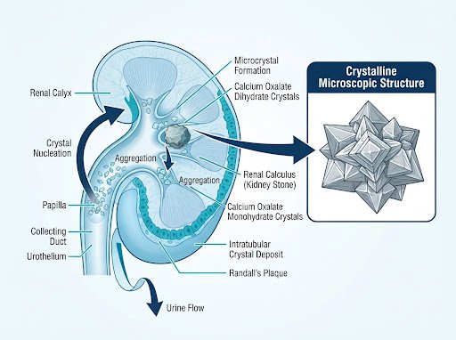
Urinary Calculus Mechanics
Minerals like calcium oxalate, phosphate, or uric acid crystallize in the renal calyx, aggregating into renal calculi that obstruct urine flow.
What Are Kidney Stones?
Kidney stones (nephrolithiasis) are hard crystalline deposits formed within the kidneys from concentrated urinary minerals. When urine contains more crystal-forming substances than the fluid can dilute, microscopic crystals aggregate into stones.
While small calculi often traverse through the ureter into the urinary bladder without causing clinical harm, larger stones can obstruct the urinary channel, causing backpressure on the kidney (hydronephrosis), severe spasmodic flank pain, and potential secondary bacterial infection.
Important Clinical Principle: Not every kidney stone requires surgical intervention. The decision to observe or operate depends entirely on size, location, symptoms, infection risk, and preservation of renal parenchyma.
How Do Kidney Stones Feel?
Kidney stone pain is widely described as one of the most intense bodily sensations. Symptoms often manifest abruptly as the calculus moves into the narrow ureteral passage.
Severe Side or Back Pain
Classic renal colic manifesting as intense, sharp cramping pain beneath the ribs, which fluctuates in severity in waves.
Pain Radiating Toward Groin
As the calculus shifts downward along the ureter, the pain pattern typically radiates into the lower abdomen, groin, or labia/testicles.
Blood in Urine (Hematuria)
Urine appears pink, reddish, or brownish due to mucosal abrasions inside the ureter or renal pelvis as the calculus shifts.
Burning During Urination
Dysuria or irritation when the stone approaches the ureterovesical junction entering the bladder cavity.
Nausea and Vomiting
Systemic autonomic reflexes triggered by the shared nerve plexus between the kidneys and gastrointestinal system.
Urinary Urgency & Frequency
Persistent urge to pass small volumes of urine repeatedly, frequently confused with simple bacterial cystitis.
Urological Red Alert: Immediate Evaluation Required
Seek urgent emergency attention if flank or back pain is accompanied by fever, shaking chills, persistent vomiting, anuria (inability to pass urine), or a single functioning kidney. Obstructive pyelonephritis is a urological emergency.
When Should You See a Urologist?
Early specialized consultation prevents silent irreversible loss of renal function and chronic kidney damage.
Persistent Colic Pain
Pain that does not subside with standard analgesics or recurs across several days.
Stone Not Passing
Calculi >5-6mm that fail to progress through the ureter after 2 to 4 weeks of medical therapy.
Urinary Obstruction
Ultrasound shows moderate or severe hydronephrosis with renal parenchymal swelling.
Frank Hematuria
Noticeable macroscopic blood or blood clots passed continuously during micturition.
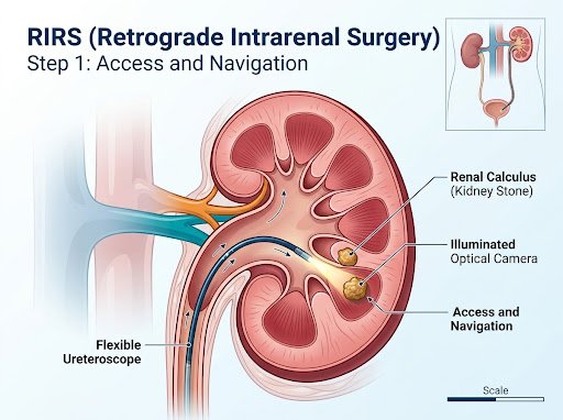
Locate & Access
Ultra-thin digital video ureteroscope navigates through the natural urethral channel into the kidney calyx to establish direct high-definition visualization of the stone.
- No cuts or incisions
- Active 270° scope deflection
- Access to lower pole calyces
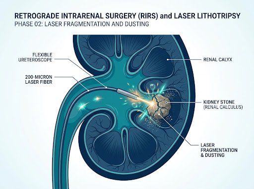
Laser Fragmentation
A 200-micron optical Holmium or Thulium laser fiber delivers high-frequency photothermal energy pulses, vaporizing the calculus into fine sub-millimeter sand particles.
- Holmium / Thulium laser fiber
- Dusting & pop-dusting mode
- Zero thermal collateral injury
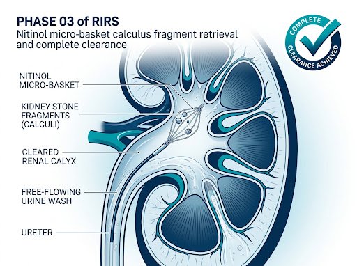
Clearance & Relief
Nitinol micro-baskets safely retrieve remaining stone fragments while microscopic dust washes out spontaneously during normal urine flow.
- Zero-residual stone clearance
- Nitinol tipless basket retrieval
- Same-day discharge recovery
How Are Kidney Stones Diagnosed?
A methodical diagnostic sequence accurately pinpoints stone dimensions, exact tract location, internal composition, and overall renal status.
Locate & Access
In-depth clinical exploration of pain trajectory, prior stone formation history, fluid habits, family risks, and medication usage.
Urinalysis & Culture
Checks for microscopic hematuria, crystalline precipitation, pyuria (white cells), and active bacterial urinary pathogens.
Renal Blood Panels
Serum creatinine and eGFR to verify filtering capacity, plus serum calcium, uric acid, and systemic inflammatory markers.
Imaging: NCCT / USG
Non-Contrast Computed Tomography (NCCT KUB) is the gold standard for definitive 3D stone localization and Hounsfield density mapping.
Clinical Practice Note: The choice of diagnostic investigation depends closely on presenting symptoms, age, pregnancy status, and prior radiation exposure considerations.
Which Treatment Is Right for a Kidney Stone?
Urological therapy is customized to the patient's individual calculus parameters, anatomical variations, and health status.
Observation & Medical Expulsive Therapy (MET)
For small asymptomatic calculi (<4-5mm) without high obstruction or severe discomfort.
- Hydration regime (>2.5-3L/day)
- Alpha-blocker smooth muscle relaxants
- Structured ultrasound follow-up
Extracorporeal Shock Wave (ESWL)
Non-invasive acoustic energy targeted under X-ray or ultrasound for select small upper renal calculi.
- Suitable for soft, <1cm stones
- Completely external sound waves
- Fragments pass naturally in urine
Ureteroscopy (URS) & RIRS Laser Surgery
Endoscopic navigation through natural urinary passages for mid-to-lower ureteric and intra-renal calculi.
- Zero external skin incisions
- Holmium / Thulium fiber dusting
- High single-stage stone clearance
Mini-PCNL (Keyhole Surgery)
For larger (>1.5–2 cm), staghorn, or complex lower-pole calyceal stones requiring direct puncture.
- Ultra-fine mini-tract (11–14 Fr)
- Fluoroscopic / Sonographic precision
- Direct vacuum / basket evacuation
- Decision Factors: Stone size, exact calyx location, radiological density (Hounsfield units), pelvic anatomy, systemic infection markers, and baseline creatinine determine the clinical path.
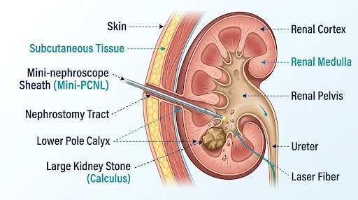
Mini-PCNL (Miniature Percutaneous Nephrolithotomy)
When kidney stones exceed 1.5 to 2 centimeters, or form dense complex staghorn configurations in the lower renal pole, Mini-PCNL is the preferred surgical procedure.
Unlike traditional open surgery or standard large-bore PCNL, Mini-PCNL utilizes an ultra-fine puncture tract (11 to 14 French—approximately 3 to 4 mm), significantly reducing blood loss, post-operative pain, and hospital recovery time.
Targeted Miniature Tract
3-4mm entry preserves renal tissue.
Ultrasound Guidance
Real-time acoustic tract puncture.
High Clearance
Complete removal of large burdens.
Mini-PCNL (Miniature Percutaneous Nephrolithotomy)
When kidney stones exceed 1.5 to 2 centimeters, or form dense complex staghorn configurations in the lower renal pole, Mini-PCNL is the preferred surgical procedure.
Unlike traditional open surgery or standard large-bore PCNL, Mini-PCNL utilizes an ultra-fine puncture tract (11 to 14 French—approximately 3 to 4 mm), significantly reducing blood loss, post-operative pain, and hospital recovery time.
Targeted Miniature Tract
3-4mm entry preserves renal tissue.
Ultrasound Guidance
Real-time acoustic tract puncture.
High Clearance
Complete removal of large burdens.
When Is Mini-PCNL Indicated?
- Calculi >15mm: Where retrograde scopes may have lower single-sitting clearance.
- Lower Pole Stones: Stones trapped in steep infundibulopelvic angles.
- Partial Staghorn Stones: Branched calculi spanning multiple renal calyces.
- Failed Prior ESWL or URS: Hard brushite or cystine stones resistant to shock waves.
Kidney Stone Treatment Modalities Compared
A balanced overview of current medical and endourological therapies. Every treatment is chosen based on your clinical safety and anatomy.
Modality
Stone Location
Invasiveness
Anaesthesia
Hospital Stay
Suitability
Medical / Observation
Any small calyx or ureter
None
Outpatient
<4-5mm asymptomatic stones
ESWL (Shock Wave)
Upper renal calyx
Sedation or Local
Day-care (2-4 hours)
<10mm non-lower-pole stones
Ureteroscopy (URS)
Mid & Lower Ureter
Spinal or General
Day-care / 24 Hours
Obstructing ureteric stones
RIRS Laser Surgery
Intra-Renal / Calyces
General Anaesthesia
12 – 24 Hours
Calculi up to 1.5–2 cm, multiple calyces
Mini-PCNL
Kidney (Pelvis & Calyces)
General Anaesthesia
24 – 48 Hours
>1.5–2cm, complex or staghorn stones
*Note: This clinical matrix provides general clinical comparisons. Specific recommendations are determined following diagnostic evaluation and imaging review.
Recovery Journey After Stone Treatment
Modern minimally invasive endourology enables swift, comfortable recovery with clear post-operative milestones.
01
Immediate Recovery
Patients mobilize comfortably within hours of endourological surgery. Vital signs, fluid intake, and initial urine output are closely tracked.
02
Stent Awareness
A temporary internal Double-J (DJ) stent prevents ureteric swelling. Mild urinary urgency or slight pink-tinged urine is completely expected.
03
Stent Removal & Scan
The internal stent is smoothly removed via a quick, painless outpatient procedure under local anesthesia between day 7 and 14.
04
Metabolic Follow-Up
Stone chemical analysis and 24-hour urinary metabolic evaluation are reviewed to build a customized recurrence prevention protocol.
How Can You Reduce the Risk of Future Stones?
Nearly 50% of stone formers experience a recurrence within 5 to 7 years if preventive lifestyle modifications are not implemented.
Optimal Hydration Protocol
Maintain daily fluid intake aiming for pale, clear urine. Diluting urine solute concentration is the single most effective way to prevent crystal nucleation and stone regrowth.
- Citrate-rich lemon water
- Avoid sugary sodas & sweetened colas
Dietary Sodium Moderation
High sodium intake forces kidneys to excrete more calcium into the urine, directly binding with oxalate to form stones. Minimizing processed foods lowers hypercalciuria.
- Restrict table salt & canned snacks
- Helps normalize urinary calcium levels
Balanced Calcium & Oxalate
Counterintuitively, normal dietary calcium from dairy binds dietary oxalates in the gut before reaching kidneys. Pair calcium-rich meals when consuming oxalate foods.
- Pair dietary dairy with greens
- Moderate high-oxalate nuts & chocolate
24-Hour Urine & Stone Analysis
Comprehensive stone chemical composition analysis and 24-hour urine metabolic evaluation to identify hyperuricosuria, hypocitraturia, or cystinuria for targeted prevention.
- Tailored prescription therapy if needed
- Annual ultrasound & renal checks
RIRS — Retrograde Intrarenal Surgery
Retrograde Intrarenal Surgery (RIRS) represents a major advancement in modern endourology. Using a microscopic, flexible digital ureteroscope, Dr. Saurabh Chipde accesses the internal calyces of the kidney directly through the body's natural urinary opening.
No Skin Incisions or Stitches
The scope gently glides retrograde via the urethra, bladder, and ureter into the renal pelvis without surgical cutting.
High-Definition Digital Deflection
The endoscope tip articulates up to 270 degrees, reaching hidden stones lodged deep inside difficult lower calyces.
Advanced Laser Dusting
Laser energy pulverizes calculi into micronized dust particles that wash out naturally in urine with minimal discomfort.
RIRS Procedural Blueprint
Anaesthesia Modality:
General or Spinal Anaesthesia
Hospitalization:
Typically 12 to 24 Hours
Internal Stent (DJ Stent):
Temporary (removed in 7-14 days)
Resume Office Work:
Within 48 – 72 Hours
- Performed with Olympus Digital Flexible Ureteroscopes & High-Wattage Laser
Frequently Asked Questions
Clear, clinically verified answers to common questions about kidney stones, diagnostic procedures, and laser surgery.
1. What causes kidney stones to form in the first place?
Kidney stones develop when urine contains excessive concentrations of crystal-forming chemicals (calcium, oxalate, uric acid) relative to fluid volume. Low daily water intake, high-sodium diets, excessive animal protein, obesity, family genetics, and metabolic conditions like hyperparathyroidism all accelerate crystallization.
2. Can a kidney stone pass naturally without surgery?
Yes, stones under 4mm to 5mm frequently pass through the ureter on their own with copious hydration and Medical Expulsive Therapy (alpha-blocker medications that relax ureteral smooth muscle). Stones larger than 6mm have significantly lower spontaneous passage rates and often require procedural assistance.
3. How do I know if my back pain is a kidney stone or muscular strain?
Muscular back pain is typically continuous, tender to localized fingertip pressure, and changes with physical posture or spinal movement. Kidney stone colic arrives in excruciating waves, is unaffected by postural adjustments, radiates toward the lower abdomen or groin, and is frequently accompanied by urinary burning, pink urine, or nausea.
4. What is the difference between RIRS and Mini-PCNL?
RIRS (Retrograde Intrarenal Surgery) enters entirely through the natural urinary channel without any skin incision, making it ideal for stones up to 1.5–2cm. Mini-PCNL makes a small 3–4mm keyhole puncture in the back under imaging guidance directly into the kidney, making it superior for larger (>2cm), multiple, or complex lower-pole calculi.
5. Is laser stone surgery painful?
The procedure itself is performed under general or regional anaesthesia, so you feel nothing during surgery. Post-operatively, mild discomfort, temporary urinary burning, and urgency may occur due to the presence of the internal stent, but these symptoms are easily managed with prescribed oral medications.
6. Why is a Double-J (DJ) stent required after laser surgery?
Manipulating instruments inside the narrow ureter creates temporary mucosal edema (swelling). The DJ stent maintains an open channel, ensuring unhindered urine drainage, preventing severe colic, and helping fine residual stone dust pass freely out of the kidney.
7. How long does the DJ stent stay in place and how is it removed?
In most standard laser lithotripsy cases, the stent remains in place for 7 to 14 days. It is subsequently removed as a simple, 2-minute outpatient clinic procedure using local anaesthetic jelly and a fine flexible cystoscope.
8. When can I return to work and routine physical activities?
Most patients undergoing RIRS or URS return to desk work within 48 to 72 hours. For Mini-PCNL, full recovery usually takes 4 to 6 days. Vigorous gym workouts or heavy weight lifting should be paused until the internal stent has been removed.
9. Is a CT scan really necessary if an ultrasound already found a stone?
While ultrasound is safe and radiation-free, it can overestimate stone dimensions by 20-30% and frequently misses stones hiding in the mid-ureter behind bowel gas. A low-dose Non-Contrast CT KUB gives the exact millimeter size, density (Hounsfield units), and 3D road map required to safely plan laser surgery.
10. Can drinking beer or barley water dissolve kidney stones?
This is a widespread and dangerous misconception. Alcohol acts as a dehydrating diuretic that can worsen hyperuricemia and accelerate stone formation. Barley water is hydrating, but no beverage dissolves solid calcium oxalate stones once formed. Ample pure water remains the medical gold standard.
11. What is the risk of kidney stones recurring in the future?
Without targeted dietary correction and adequate hydration, recurrence rates approximate 30% to 50% over a 5-year span. Under Dr. Chipde's structured metabolic prevention protocols, this recurrence risk is substantially lowered.
12. Does health insurance cover laser kidney stone surgeries?
Yes, therapeutic endourological procedures like RIRS, URS, and Mini-PCNL are recognized medical necessities and are routinely covered under cashless mediclaim policies and corporate health coverage at Rajshree Apollo Hospitals, Indore.
Don't Know Which Stone Treatment You Need?
Bring your Ultrasound, KUB X-Ray, or CT Scan report for a personalized evaluation with Dr. Saurabh Sudhir Chipde. We'll map the safest, least invasive path for complete stone clearance.
Visit Dr. Chipde at Rajshree Apollo Hospitals Indore
Conveniently accessible in Vijay Nagar, Indore. Book an express in-clinic consultation or connect on WhatsApp.
Rajshree Apollo Hospitals, Indore
- Practice Address:
Rajshree Apollo Hospitals, Sector-D, Scheme No. 74C, Vijay Nagar, Indore, Madhya Pradesh 452010
- OPD Clinic Hours:
Monday to Saturday: 10:00 AM – 06:00 PM (Sunday by emergency appointment)
- Direct Phone & WhatsApp: